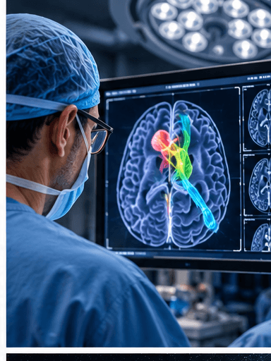
At the base of the brain, a few millimeters can separate a tumor from blood vessels, the pituitary gland and nerves that control vision.
Surgeons already bring extensive training, imaging and judgment to that environment. Researchers at University College London are testing whether artificial intelligence can provide another useful layer of information during the operation itself.
At the National Hospital for Neurology and Neurosurgery, the first patient entered a clinical trial of a UCL-developed system during surgery to remove a pituitary tumor.
The AI analyzed the live endoscopic video feed rather than relying only on scans taken before surgery. It highlighted critical anatomical structures and risky areas for the surgical team to avoid.
The system did not operate independently or make the final decisions. The surgeon remained in control throughout the procedure.
The tumor was successfully removed, the patient's vision was protected and UCL reported that it subsequently improved.
Researchers trained and evaluated the system using annotated videos from earlier pituitary operations. The ongoing trial is intended to assess whether it can assist surgeons safely and usefully in real time.
This is a more grounded form of medical AI than a general promise about what algorithms may someday do: a specific tool was used during a real procedure for a defined task.
The next stage is measurement. Researchers still need to report feasibility, safety and clinical outcomes across more patients before anyone can know whether the approach reliably improves care.